
Navigating the complexities of head and neck tumors requires a deep understanding of both the disease process and the intricate anatomy of the affected regions. For many patients, the initial symptoms may seem innocuous—a persistent sore throat, a mouth ulcer that refuses to heal, or a subtle change in voice. However, early detection and prompt intervention by a dedicated head and neck specialist can dramatically alter the course of the disease. The philosophy of modern surgical oncology dictates that removing the cancer is only half the battle; restoring the patient's form and function is equally imperative. This dual objective has given rise to the specialized field of microvascular reconstructive surgery, which works hand-in-hand with tumor resection. As we delve into the specifics of treatment, it becomes evident that a multidisciplinary approach, combining the skills of surgeons, radiation oncologists, and medical oncologists, provides the most robust defense against these aggressive malignancies.
The Complex Structures of the Head and Neck
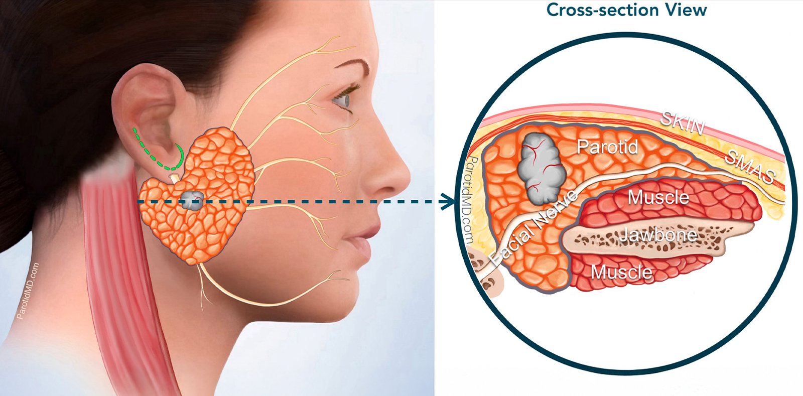
To fully appreciate the intricacies of head and neck surgery, one must understand the dense concentration of vital structures in this relatively small anatomical area. The neck houses major blood vessels, including the carotid arteries and jugular veins, which supply and drain the brain. It also contains critical nerves, such as the vagus nerve, the hypoglossal nerve (controlling tongue movement), and the facial nerve. When a tumor develops in the parotid gland, for example, it often wraps around the facial nerve, making its surgical removal an exercise in extreme precision to avoid facial paralysis. Furthermore, the lymphatic system in the neck, consisting of hundreds of lymph nodes divided into different levels, acts as the primary pathway for the metastatic spread of head and neck cancers. A comprehensive surgical plan almost always includes a neck dissection—the systematic removal of these lymph nodes—to stage the disease accurately and prevent regional recurrence.
Understanding the Etiology of the Disease
While tobacco and alcohol remain the most significant culprits in the development of head and neck malignancies, several other risk factors contribute to the disease burden. Poor oral hygiene and chronic dental trauma, such as ill-fitting dentures or sharp, jagged teeth that constantly rub against the tongue or cheek lining, can induce chronic inflammation. Over time, this constant cellular irritation can trigger mutations that lead to cancer. Nutritional deficiencies, particularly a lack of fresh fruits and vegetables rich in antioxidants, can weaken the body's natural defense mechanisms against cellular damage. Occupational exposures to certain dusts and chemicals, such as wood dust, nickel dust, and formaldehyde, are known risk factors for cancers of the paranasal sinuses and nasal cavity. Additionally, genetic predispositions and inherited syndromes, though rare, can increase an individual's susceptibility. Understanding these risk factors is not only crucial for prevention but also helps the surgical oncologist tailor the screening and diagnostic process for high-risk individuals.
The Path to an Accurate Diagnosis
Recognizing the early symptoms of head and neck cancer can mean the difference between a minor surgical procedure and a massive, life-altering operation. Patients should be vigilant about any unexplained numbness in the facial or oral regions, loosening of teeth without apparent dental disease, or a persistent earache that does not respond to standard treatments (which can often be referred pain from a tumor in the throat). The diagnostic journey typically begins with a thorough clinical examination, including a fiber-optic endoscopy to visualize the hidden areas of the throat and larynx. Following this, a targeted biopsy is performed. For neck masses, a Fine Needle Aspiration Cytology (FNAC) is a quick and relatively painless way to obtain cells for analysis. Radiographic evaluation is indispensable for surgical planning. High-resolution MRI is particularly useful for assessing soft tissue involvement and perineural spread (cancer tracking along nerves), while a CT scan is excellent for evaluating bone erosion, such as tumor invasion into the mandible (lower jaw) or maxilla (upper jaw).
Advanced Oncologic Surgery Techniques
Modern head and neck surgery has evolved to maximize tumor eradication while minimizing collateral damage to the patient. For tumors located in the deeper regions of the throat, such as the oropharynx or larynx, traditional open surgeries often required splitting the jaw or making large neck incisions. Today, selected cases can be treated using Transoral Robotic Surgery (TORS) or Transoral Laser Microsurgery (TLM). These minimally invasive techniques allow the surgeon to access and remove tumors through the mouth using high-definition 3D cameras and articulating robotic arms, leading to dramatically reduced recovery times, less scarring, and better preservation of swallowing and speech functions. However, for large, invasive tumors of the oral cavity and facial bones, open surgery remains the gold standard. In these complex cases, the surgical oncologist employs a 3D virtual surgical planning system, using computer models of the patient's anatomy to precisely map out the bone cuts and the reconstructive plan before the patient even enters the operating room.
Restoring Form and Function
The integration of microvascular reconstructive surgery with oncologic resection is what defines a world-class head and neck cancer center. The reconstructive process is tailored entirely to the specific defect created by the tumor removal. If a massive portion of the tongue is resected (total or subtotal glossectomy), an Anterolateral Thigh (ALT) flap or a Radial Forearm Free Flap (RAFF) is used to create a new, bulky neo-tongue, enabling the patient to swallow and articulate speech. When the upper jaw (maxilla) or lower jaw (mandible) is removed, vascularized bone flaps (like the fibula or scapula) are sculpted to perfectly match the patient's native facial contours, often guided by custom-made 3D-printed cutting guides and titanium plates. The success of these microvascular procedures approaches 95-98% in expert hands. The profound impact of reconstruction goes beyond physical restoration; it provides immense psychological support, allowing patients to confidently reintegrate into society and maintain a high quality of life after surviving cancer.
Postoperative Care and the Road to Normalcy
Recovery from major head and neck surgery, particularly when combined with microvascular reconstruction, is a gradual process that requires a dedicated, multidisciplinary team approach. Patients typically spend the first few days in the Intensive Care Unit (ICU) for close monitoring of the reconstructed flap and airway management. Nutritional support is paramount; since patients cannot eat by mouth initially, feeding is facilitated through a nasogastric or PEG tube. As healing progresses, intensive speech and swallow therapy commence. The brain and muscles must adapt to the newly reconstructed anatomy. Physical therapy is also crucial for the recovery of the donor site (where the tissue was harvested). Consistent follow-up and adherence to rehabilitation protocols determine the ultimate functional success of the surgery. Dr. Manish S. Tiwari and his dedicated oncology team in Kolkata are committed to providing unparalleled surgical excellence and compassionate postoperative care. By combining aggressive tumor resection with state-of-the-art microvascular reconstruction, they ensure that every patient is given the best possible chance at a cancer-free life with dignity and optimal functional restoration.