
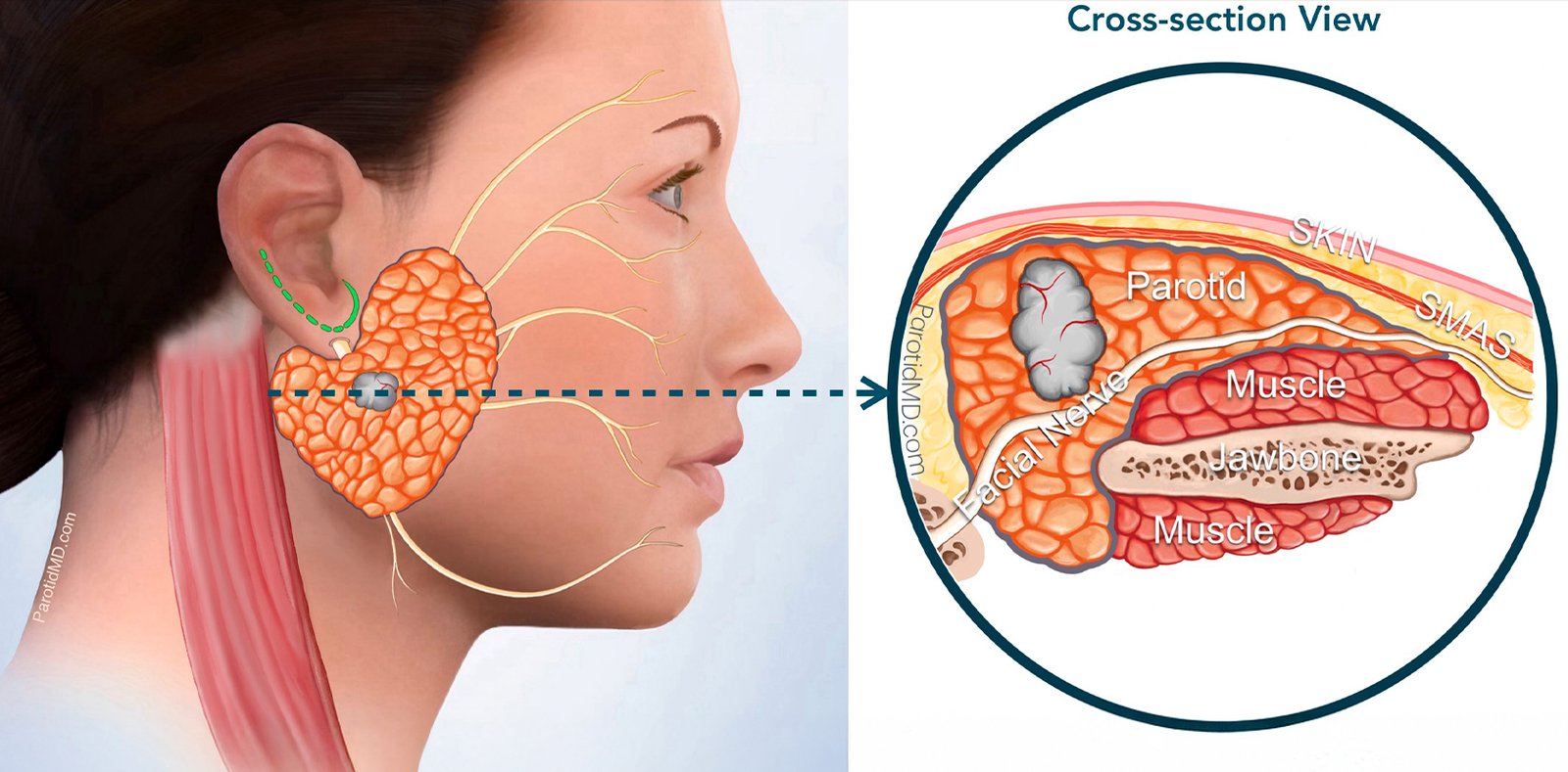
When faced with a diagnosis of cancer in the head, neck, or oral cavity, the sheer volume of information can be overwhelming. The anatomical complexity of the mouth, throat, and neck requires a highly specialized approach to treatment. Unlike other forms of cancer where surgery might be straightforward, head and neck oncology demands a delicate balance between aggressively eradicating the tumor and preserving the vital functions of speech, swallowing, and appearance. Selecting the right specialist is not just a choice; it is the most critical decision a patient and their family will make. A specialized surgical oncologist brings years of targeted experience, understanding the intricate web of nerves, blood vessels, and muscles that define the human face and neck. With the rapid advancement of modern medical technology, the paradigms of treatment have shifted significantly towards minimally invasive techniques and immediate reconstruction. Patients today have access to world-class care that prioritizes both survival and the quality of life post-treatment.
Understanding the Anatomy and Pathology
The oral cavity and the broader head and neck region consist of multiple sub-sites, each with its own unique biological behavior and lymphatic drainage patterns. The oral cavity includes the lips, the anterior two-thirds of the tongue, the buccal mucosa (inner lining of the cheeks), the floor of the mouth, the hard palate, and the upper and lower gums (alveolus). Tumors arising in different sub-sites present unique surgical challenges. For instance, a tumor on the lateral border of the tongue may spread quickly to the lymph nodes in the neck due to the tongue's rich lymphatic network. In contrast, a tumor on the hard palate might invade the maxillary sinus or nasal cavity, requiring a completely different surgical approach. Most oral cancers are squamous cell carcinomas, originating from the flat cells that line the mouth and throat. Understanding the histopathology of the tumor—its grade, depth of invasion, and perineural spread—is crucial for the surgical oncologist to plan the extent of resection and determine the necessity of postoperative radiation or chemotherapy.
Understanding the Etiology of the Disease
While tobacco and alcohol remain the most significant culprits in the development of head and neck malignancies, several other risk factors contribute to the disease burden. Poor oral hygiene and chronic dental trauma, such as ill-fitting dentures or sharp, jagged teeth that constantly rub against the tongue or cheek lining, can induce chronic inflammation. Over time, this constant cellular irritation can trigger mutations that lead to cancer. Nutritional deficiencies, particularly a lack of fresh fruits and vegetables rich in antioxidants, can weaken the body's natural defense mechanisms against cellular damage. Occupational exposures to certain dusts and chemicals, such as wood dust, nickel dust, and formaldehyde, are known risk factors for cancers of the paranasal sinuses and nasal cavity. Additionally, genetic predispositions and inherited syndromes, though rare, can increase an individual's susceptibility. Understanding these risk factors is not only crucial for prevention but also helps the surgical oncologist tailor the screening and diagnostic process for high-risk individuals.
Warning Signs and Diagnostic Modalities
Early detection remains the single most important factor in achieving a cure in head and neck oncology. Unfortunately, many patients ignore the initial warning signs, attributing them to benign conditions. Key symptoms that warrant immediate evaluation by a specialist include a persistent mouth ulcer that fails to heal within two to three weeks, unexplained bleeding in the mouth, difficulty or pain during swallowing (dysphagia), a change in voice or persistent hoarseness, and the sudden appearance of a painless lump in the neck. To accurately diagnose and stage the disease, a multifaceted approach is required. The gold standard for diagnosis is a biopsy, where a small tissue sample is extracted and examined under a microscope. Once malignancy is confirmed, advanced imaging techniques come into play. Contrast-enhanced CT scans or MRI scans provide detailed cross-sectional views of the tumor, delineating its exact size, depth of tissue invasion, and involvement of adjacent bones or blood vessels. A PET-CT scan may also be ordered to detect microscopic spread to regional lymph nodes or distant organs, ensuring that the treatment plan addresses the entirety of the disease.
Advanced Oncologic Surgery Techniques
Modern head and neck surgery has evolved to maximize tumor eradication while minimizing collateral damage to the patient. For tumors located in the deeper regions of the throat, such as the oropharynx or larynx, traditional open surgeries often required splitting the jaw or making large neck incisions. Today, selected cases can be treated using Transoral Robotic Surgery (TORS) or Transoral Laser Microsurgery (TLM). These minimally invasive techniques allow the surgeon to access and remove tumors through the mouth using high-definition 3D cameras and articulating robotic arms, leading to dramatically reduced recovery times, less scarring, and better preservation of swallowing and speech functions. However, for large, invasive tumors of the oral cavity and facial bones, open surgery remains the gold standard. In these complex cases, the surgical oncologist employs a 3D virtual surgical planning system, using computer models of the patient's anatomy to precisely map out the bone cuts and the reconstructive plan before the patient even enters the operating room.
The Miracle of Microvascular Reconstruction
Removing a large tumor from the head or neck inevitably creates a significant structural and functional deficit. Decades ago, patients were left with severe disfigurements and lifelong disabilities in eating and speaking. Today, the advent of microvascular free flap reconstructive surgery has revolutionized patient rehabilitation. This highly intricate procedure involves harvesting living tissue—skin, muscle, and bone—from a 'donor site' on the patient's own body, such as the forearm, thigh, or lower leg (fibula). The surgeon transplants this tissue to the head and neck to rebuild the missing structures. Using a high-powered operating microscope and sutures finer than a human hair, the surgeon meticulously connects the tiny arteries and veins of the transplanted tissue to the blood vessels in the neck. This restores blood flow and keeps the reconstructed tissue alive. For example, a fibula free flap can be used to completely rebuild a resected jawbone, allowing for the eventual placement of dental implants and a return to normal chewing function.
Long-Term Rehabilitation and Follow-Up
Surviving head and neck cancer is a monumental victory, but the journey continues well after the surgical incisions have healed. Comprehensive postoperative rehabilitation is vital for maximizing the functional outcomes of reconstructive surgery. Patients engage in rigorous swallowing exercises to prevent aspiration and speech therapy to improve articulation. Dietary modifications are gradually introduced as the patient transitions from tube feeding back to an oral diet. The psychological impact of facial surgery cannot be overstated, and access to counseling and support groups forms a critical component of holistic cancer care. Furthermore, rigorous surveillance is mandatory for the first five years post-treatment to monitor for any signs of recurrence. Regular clinical examinations and imaging scans provide peace of mind and ensure any issues are addressed immediately. If you or a loved one are facing a diagnosis of oral or head and neck cancer, choosing an expert who manages both the oncologic and reconstructive aspects of care is essential. Dr. Manish Tiwari represents the pinnacle of surgical expertise in Kolkata, offering hope, cutting-edge treatment, and comprehensive rehabilitation to his patients.