
A diagnosis of oral or head and neck cancer is a life-altering event that brings forth a multitude of questions and concerns. The journey from diagnosis to recovery is paved with critical decisions, the most important being the selection of an experienced surgical oncologist. The head and neck region is arguably the most complex area of the human body, housing the vital apparatus for breathing, eating, speaking, and expressing emotion. Consequently, surgical intervention in this area requires an unparalleled level of precision and expertise. Today, the field of head and neck oncology has evolved far beyond mere tumor removal. It encompasses a holistic approach where oncologic safety is seamlessly integrated with functional rehabilitation and aesthetic preservation. Advanced imaging, multidisciplinary tumor boards, and state-of-the-art reconstructive techniques have transformed the prognosis for many patients. Understanding the nuances of the disease and the available treatment modalities empowers patients to take an active role in their healthcare journey, leading to better psychological and physical outcomes.
Understanding the Anatomy and Pathology
The oral cavity and the broader head and neck region consist of multiple sub-sites, each with its own unique biological behavior and lymphatic drainage patterns. The oral cavity includes the lips, the anterior two-thirds of the tongue, the buccal mucosa (inner lining of the cheeks), the floor of the mouth, the hard palate, and the upper and lower gums (alveolus). Tumors arising in different sub-sites present unique surgical challenges. For instance, a tumor on the lateral border of the tongue may spread quickly to the lymph nodes in the neck due to the tongue's rich lymphatic network. In contrast, a tumor on the hard palate might invade the maxillary sinus or nasal cavity, requiring a completely different surgical approach. Most oral cancers are squamous cell carcinomas, originating from the flat cells that line the mouth and throat. Understanding the histopathology of the tumor—its grade, depth of invasion, and perineural spread—is crucial for the surgical oncologist to plan the extent of resection and determine the necessity of postoperative radiation or chemotherapy.
Understanding the Etiology of the Disease
While tobacco and alcohol remain the most significant culprits in the development of head and neck malignancies, several other risk factors contribute to the disease burden. Poor oral hygiene and chronic dental trauma, such as ill-fitting dentures or sharp, jagged teeth that constantly rub against the tongue or cheek lining, can induce chronic inflammation. Over time, this constant cellular irritation can trigger mutations that lead to cancer. Nutritional deficiencies, particularly a lack of fresh fruits and vegetables rich in antioxidants, can weaken the body's natural defense mechanisms against cellular damage. Occupational exposures to certain dusts and chemicals, such as wood dust, nickel dust, and formaldehyde, are known risk factors for cancers of the paranasal sinuses and nasal cavity. Additionally, genetic predispositions and inherited syndromes, though rare, can increase an individual's susceptibility. Understanding these risk factors is not only crucial for prevention but also helps the surgical oncologist tailor the screening and diagnostic process for high-risk individuals.
Warning Signs and Diagnostic Modalities
Early detection remains the single most important factor in achieving a cure in head and neck oncology. Unfortunately, many patients ignore the initial warning signs, attributing them to benign conditions. Key symptoms that warrant immediate evaluation by a specialist include a persistent mouth ulcer that fails to heal within two to three weeks, unexplained bleeding in the mouth, difficulty or pain during swallowing (dysphagia), a change in voice or persistent hoarseness, and the sudden appearance of a painless lump in the neck. To accurately diagnose and stage the disease, a multifaceted approach is required. The gold standard for diagnosis is a biopsy, where a small tissue sample is extracted and examined under a microscope. Once malignancy is confirmed, advanced imaging techniques come into play. Contrast-enhanced CT scans or MRI scans provide detailed cross-sectional views of the tumor, delineating its exact size, depth of tissue invasion, and involvement of adjacent bones or blood vessels. A PET-CT scan may also be ordered to detect microscopic spread to regional lymph nodes or distant organs, ensuring that the treatment plan addresses the entirety of the disease.
Surgical Interventions and Treatment Strategies
The cornerstone of curative treatment for early and moderately advanced oral cancers is surgical resection. The primary objective is to achieve 'negative margins'—removing the tumor entirely along with a surrounding cuff of healthy tissue to ensure no microscopic cancer cells are left behind. Depending on the tumor's location, procedures can range from a simple wide local excision of a tongue lesion (partial glossectomy) to a massive composite resection that includes the removal of a portion of the jawbone (mandibulectomy or maxillectomy). Because oral cancers frequently metastasize to the lymph nodes in the neck, a 'neck dissection' is concurrently performed. This meticulous procedure involves carefully stripping the fat and lymph nodes from the vital nerves and blood vessels of the neck. For advanced cases, surgery is often just the first step in a multimodal treatment plan. Based on the final pathology report, the patient may require adjuvant (post-operative) radiation therapy or concurrent chemo-radiation to eradicate any lingering microscopic disease and significantly reduce the risk of recurrence.
The Miracle of Microvascular Reconstruction
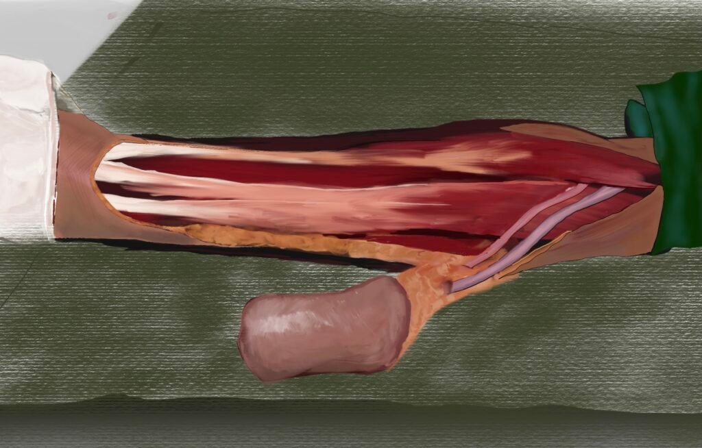
Removing a large tumor from the head or neck inevitably creates a significant structural and functional deficit. Decades ago, patients were left with severe disfigurements and lifelong disabilities in eating and speaking. Today, the advent of microvascular free flap reconstructive surgery has revolutionized patient rehabilitation. This highly intricate procedure involves harvesting living tissue—skin, muscle, and bone—from a 'donor site' on the patient's own body, such as the forearm, thigh, or lower leg (fibula). The surgeon transplants this tissue to the head and neck to rebuild the missing structures. Using a high-powered operating microscope and sutures finer than a human hair, the surgeon meticulously connects the tiny arteries and veins of the transplanted tissue to the blood vessels in the neck. This restores blood flow and keeps the reconstructed tissue alive. For example, a fibula free flap can be used to completely rebuild a resected jawbone, allowing for the eventual placement of dental implants and a return to normal chewing function.
Postoperative Care and the Road to Normalcy
Recovery from major head and neck surgery, particularly when combined with microvascular reconstruction, is a gradual process that requires a dedicated, multidisciplinary team approach. Patients typically spend the first few days in the Intensive Care Unit (ICU) for close monitoring of the reconstructed flap and airway management. Nutritional support is paramount; since patients cannot eat by mouth initially, feeding is facilitated through a nasogastric or PEG tube. As healing progresses, intensive speech and swallow therapy commence. The brain and muscles must adapt to the newly reconstructed anatomy. Physical therapy is also crucial for the recovery of the donor site (where the tissue was harvested). Consistent follow-up and adherence to rehabilitation protocols determine the ultimate functional success of the surgery. Dr. Manish S. Tiwari and his dedicated oncology team in Kolkata are committed to providing unparalleled surgical excellence and compassionate postoperative care. By combining aggressive tumor resection with state-of-the-art microvascular reconstruction, they ensure that every patient is given the best possible chance at a cancer-free life with dignity and optimal functional restoration.